8 min read
Iodine Pineal Gland: The NIS Mechanism 4M Sites Got Wrong
4 million results claim iodine decalcifies the pineal gland. The NIS mechanism is real — the extrapolation isn't. Here's where the evidence...
April 14, 2026
Read →
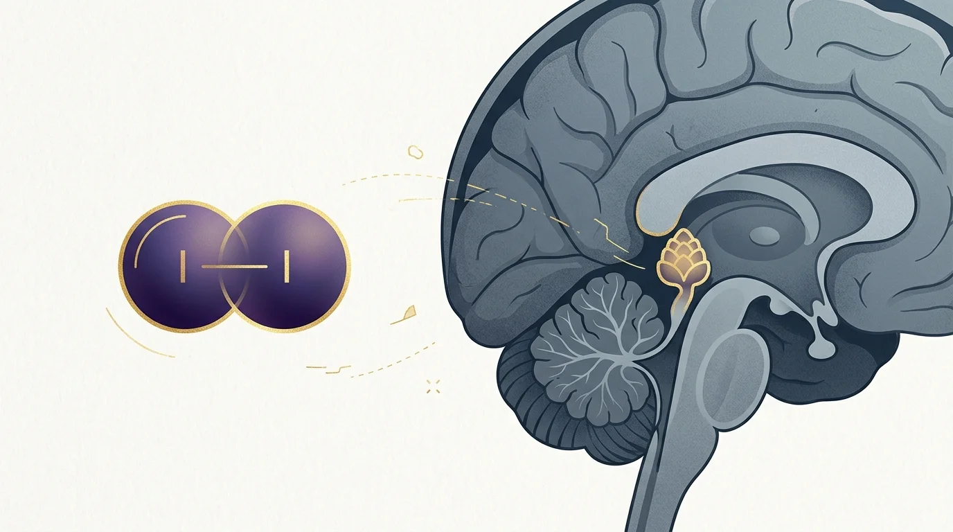
Your pineal gland contains actual crystals. Not metaphorical crystals. Not “crystal energy.” Physical, measurable, peer-reviewed crystals — sitting inside a pea-sized gland in the center of your brain right now.
Science has known this since at least 2002, when researchers at Ben-Gurion University published the first detailed physical and chemical study of pineal microcrystals in Bioelectromagnetics. And yet the conversation around “pineal gland crystals” is almost entirely dominated by either mystical speculation or reflexive debunking.
Both miss what’s actually interesting.
There are two distinct types of crystals in the pineal gland — with different compositions, different origins, and very different implications for your health. One is a sign of something going wrong. The other may have been there since you were born. Understanding the difference matters — especially if you’ve been reading about pineal calcification symptoms and wondering what they mean for you.
Here’s what the research actually shows.
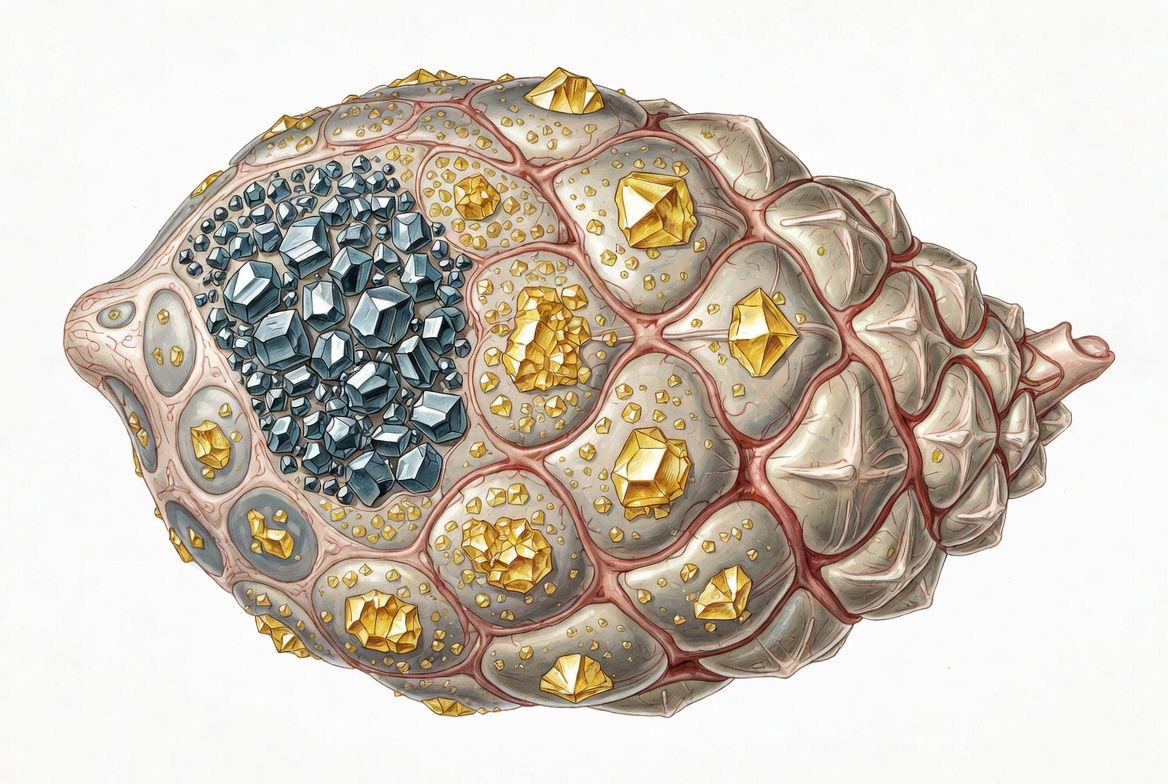
Yes — and they’ve been documented in two chemically distinct forms.
The first are calcite microcrystals (calcium carbonate, CaCO₃), identified in all 20 human pineal specimens examined in Baconnier and Lang’s 2002 study in Bioelectromagnetics.
The second are magnetite crystals (iron oxide, Fe₃O₄), first confirmed in human brain tissue by Kirschvink and colleagues in a 1992 PNAS paper using a superconducting magnetometer.
These are not the same thing. They don’t form the same way. Conflating them — which nearly every wellness article does — produces exactly the kind of confusion that leads people to panic about something natural, or brush off something worth paying attention to.
The calcite crystals appeared as cubic, hexagonal, and cylindrical structures, all smaller than 20 micrometers. The researchers noted their structural similarity to otoconia — the calcium carbonate crystals in your inner ear that help you detect movement and gravity. That similarity has real implications, which I’ll get to.
Kirschvink’s team found a minimum of 5 million single-domain magnetite crystals per gram in most brain tissue, and more than 100 million per gram in the meninges. The pineal gland wasn’t the specific focus of that study, so precision localization in the pineal is still an open question — but the presence of magnetite in human neural tissue is not in dispute.
For a deeper look at what those piezoelectric properties might mean, the pineal gland piezoelectricity article covers the Baconnier findings in full detail.
The answer depends entirely on which crystals you’re asking about — and this is where almost every popular article gets it wrong.
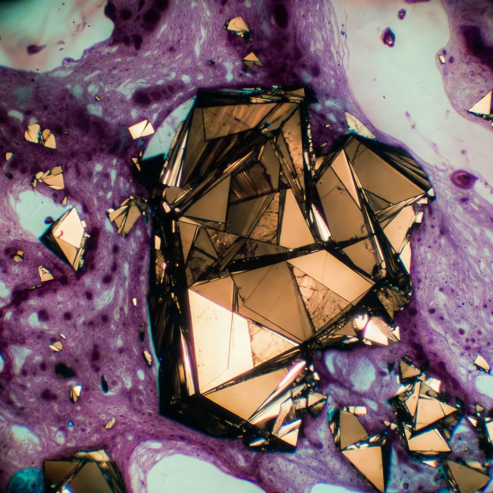
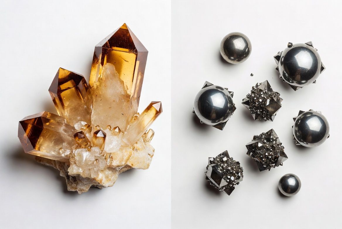
The calcite microcrystals Baconnier identified are composed of calcium carbonate — the same mineral compound that forms limestone, chalk, and the balance-sensing otoconia in your inner ear. This is not the same as hydroxyapatite, which is the calcium phosphate compound associated with standard pathological calcification elsewhere in the body.
Calcite microcrystals in the human pineal gland — composed of CaCO₃, not the hydroxyapatite of ordinary calcification — were found in 100% of the 20 specimens examined by Baconnier et al. (2002), at densities of 100–300 crystals per cubic millimeter, with structural properties similar to piezoelectric transducers in the inner ear.
That distinction matters. The pineal gland appears to produce a structurally specific form of crystal — not just generic calcium buildup. Why? The original researchers flagged it as an area for further investigation. Meaningful follow-up has been scarce, which is either reassuring or frustrating depending on how you look at it.
Magnetite is a naturally magnetic iron oxide. In brain tissue, it’s not considered pathological — the current thinking is that the body synthesizes it deliberately. The leading hypothesis is that it acts as a kind of biological compass, providing sensitivity to geomagnetic fields. Similar mechanisms have been demonstrated in migratory birds.
That hypothesis hasn’t been experimentally confirmed in humans. The crystals are real, present from early development, and there’s no evidence they cause harm.
No — and this is the most important distinction in this entire article.
When most people say “pineal calcification,” they mean calcium deposits accumulating in the gland’s tissue over time, visible as a bright spot on CT scans. This involves hydroxyapatite — calcium phosphate — and is associated with reduced melatonin output and disrupted sleep.
The calcite microcrystals Baconnier described are chemically distinct from that process. They’re smaller, structurally different, and present in essentially everyone — not increasing progressively with age the way pathological calcification does.
Two chemically distinct phenomena — one natural, one pathological
The magnetite crystals belong in the “natural and universal” column as well.
So when someone says “pineal crystals are bad,” they’re almost certainly talking about calcification — not the microcrystals Baconnier found. Both exist. They’re not the same. Treating them as interchangeable produces bad conclusions.
Honest answer: we don’t fully know. The Baconnier study identified their structure and composition but didn’t establish the mechanism by which the pineal gland synthesizes calcium carbonate crystals instead of — or alongside — ordinary hydroxyapatite deposits. It’s one of the genuine open questions in this research area, and I find it a bit odd that more labs haven’t tried to answer it.
This process is better understood. Calcium phosphate deposits accumulate gradually with age, influenced by fluoride exposure, calcium metabolism, and diet. Luke’s 2001 study finding fluoride concentrations around 9,000 mg/kg in pineal hydroxyapatite — published in Caries Research — found that fluoride concentrates at levels exceeding those found in bone, with a strong positive correlation between fluoride and calcium levels in the gland (r=0.73).
I used to think the fluoride-pineal connection was mostly overstated by people selling supplements. Then I read Luke’s data. It’s not as dramatic as the wellness crowd makes it sound — but the accumulation is real, and the correlation with calcium is hard to dismiss.
Calcification becomes detectable on imaging in many people by their twenties or thirties. A 2023 systematic review and meta-analysis found a pooled prevalence of roughly 62% in adults, ranging from 35% in some regions to 76% in others, with no significant difference between sexes.
Magnetite appears to be present from fetal development or early childhood. The body synthesizes it. No environmental trigger has been identified, no intervention is needed, and if pineal magnetite does serve a function — still speculative — interfering with it would presumably be counterproductive.
This is where things get genuinely strange — in a scientifically respectable way.
Calcite is a piezoelectric material: it generates an electrical charge under mechanical pressure. Baconnier’s team observed second harmonic generation (SHG) in pineal tissue sections — an optical phenomenon tied to piezoelectric materials — and proposed the calcite microcrystals could function as piezoelectric transducers.
The analogy they drew was to otoconia — the calcium carbonate crystals in the inner ear that convert motion into electrical signals your brain reads as position and movement. If pineal calcite works similarly, it could theoretically convert mechanical or electromagnetic signals into electrical impulses within the gland.
Theoretically. No study has demonstrated this in living human tissue. The piezoelectric hypothesis is structurally plausible and supported by the SHG data — but unconfirmed in vivo. Whether it ever gets confirmed depends largely on whether anyone bothers to fund the research.
The full breakdown is in the pineal gland piezoelectricity article. And for the specific question of whether WiFi or residential EMFs can interact with these crystals in a clinically meaningful way, the EMF and pineal gland research separates what’s established from what remains hypothesis.
Calcification does. Magnetite crystals don’t appear to.
A 1999 study by Kunz et al. linking uncalcified pineal tissue volume to melatonin metabolite excretion in 26 subjects established the core relationship: more calcification correlated with less melatonin. A 2009 study by Mahlberg et al. in Sleep Medicine extended this to clinical outcomes: in 31 patients with primary insomnia, greater calcification severity was inversely associated with REM sleep, total sleep time, and sleep efficiency.
Both studies are small. Neither establishes causation — we don’t know if calcification destroys pinealocytes, compresses them, or interferes with signaling through some other mechanism. But the correlation between calcification and disrupted sleep architecture is consistent enough across studies that I take it seriously.
If you’re already reading about decalcification at night protocols, the Kunz and Mahlberg findings are the scientific scaffolding for why that matters.
The calcite microcrystals Baconnier identified? You wouldn’t want to — and there’s no known way to do so anyway. They appear to be a normal feature of pineal anatomy. Same with magnetite. Leave it alone.
Calcification (hydroxyapatite deposits) is the one worth attention — and there’s indirect evidence it can be slowed or partially reversed, though no randomized clinical trial has tested any intervention specifically in pineal tissue.
What the indirect evidence supports:
These are preventive and support strategies backed by indirect evidence — not proven cures for established calcification. Anyone presenting them as guaranteed reversals is overstating the research.
For the complete breakdown on protocols and methods, how to decalcify the pineal gland naturally covers what actually has support behind it.
And if you’re looking at supplements specifically, the best supplements for pineal decalcification guide breaks down what works.
Two types of crystals. Two completely different stories.
The calcite microcrystals and magnetite are almost certainly in your pineal gland right now. They’re not pathological. They’re not a problem. Whether they do anything interesting — piezoelectric transduction, magnetic sensitivity — is a genuinely open scientific question that deserves more attention than it gets.
Calcification is the real concern. It builds with age, appears to reduce melatonin and disrupt sleep, and accumulates fluoride in a way that’s hard to ignore. It’s also something you have partial control over — especially if you start before it’s advanced.
The crystals that belong there: don’t worry about them. The deposits that accumulate over decades: that’s where your attention should go.
Start with sleep quality. See if it shifts. And if you want to go further, read the calcification symptoms piece to understand what compromised pineal function actually looks like.
Marcus Hale is an independent researcher and former clinical neuroscientist. The content on PinealCode.com is for informational purposes only and does not constitute medical advice.
4 million results claim iodine decalcifies the pineal gland. The NIS mechanism is real — the extrapolation isn't. Here's where the evidence...
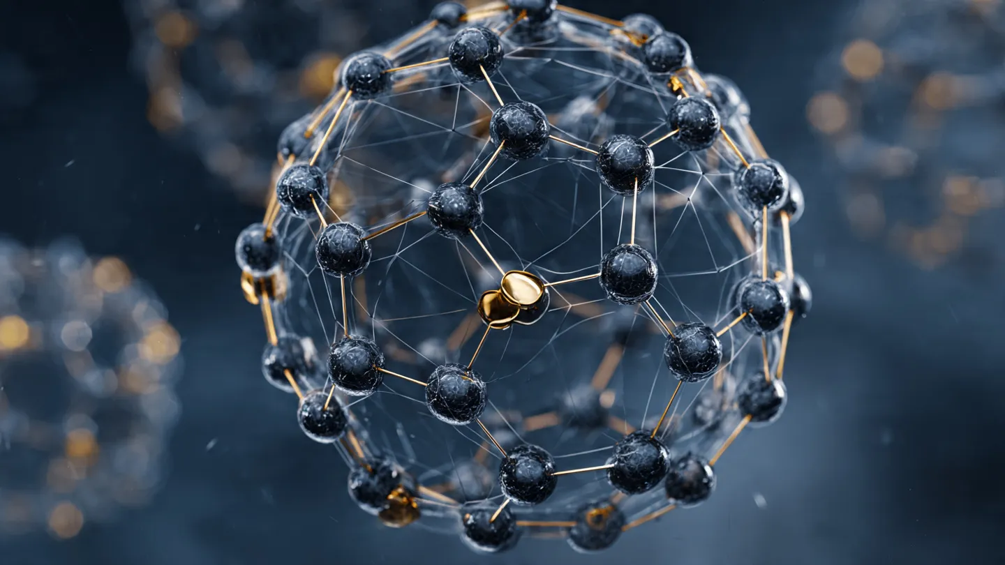
C60 binds up to 6 electrons at once — no other antioxidant works this way. Whether it protects your pineal from calcification: the honest...
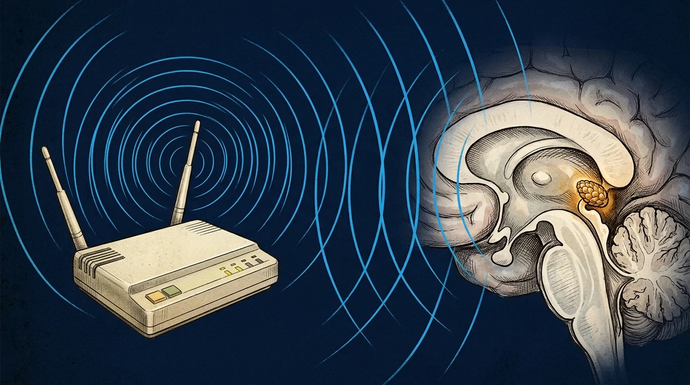
WiFi and pineal calcification — what the science actually shows. Volkow 2011, Baconnier 2002 crystals, calcium channels. Honest analysis,...

Marcus Hale
Independent Researcher · Former Clinical Neuroscientist
I spent 12 years in clinical neurology before the questions got more interesting than the answers. PinealCode is where I document what I find at the intersection of brain science and consciousness.