8 min read
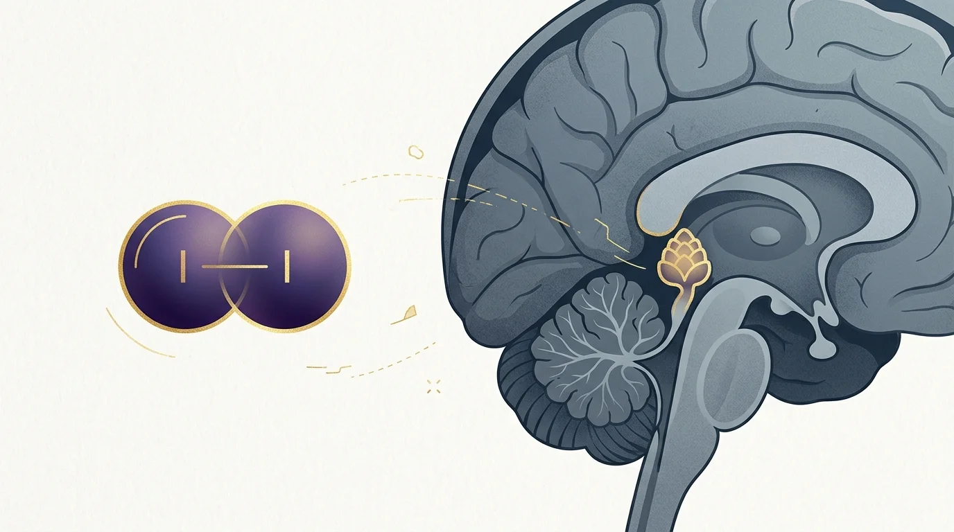
Iodine Pineal Gland: The NIS Mechanism 4M Sites Got Wrong
4 million results claim iodine decalcifies the pineal gland. The NIS mechanism is real — the extrapolation isn't. Here's where the evidence...
April 14, 2026
Read →
Something real was found in 2002.
A team of researchers published a paper in Bioelectromagnetics announcing the discovery of calcite microcrystals in the human pineal gland — a form of biomineralization that had never been documented there before. Not the calcium phosphate crust that builds up with age. Something different. Smaller. Structurally precise. And composed of a mineral with documented piezoelectric properties.
Then almost nothing happened. For over twenty years.
That’s the story I want to tell here — not the spiritual version you’ve probably already read, and not the dismissive debunking either. The science is real. The gap that followed is strange. And the line between what was found and what was claimed deserves a cleaner map than this niche usually draws.
Yes and no — and the distinction between those two answers is the entire point of this article.
The pineal gland contains calcite microcrystals, and calcite has documented piezoelectric properties in mineralogy. That part is real. What has not been demonstrated — in any published study, in any model, in any living tissue — is whether the pineal gland as a functioning biological system generates or responds to electromagnetic fields via those crystals.
Those are two different claims. The first is anatomy. The second is functional physiology. Most of the confusion in this space comes from running them together.
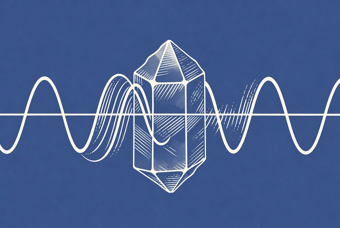
Piezo comes from the Greek for “press.” Compress certain crystals mechanically and they generate voltage. Apply an electric field to those same crystals and they deform or vibrate. That’s it.
It’s not speculative. It’s not fringe. Your gas kitchen lighter runs on it. Medical ultrasound runs on it. Contact microphones, sonar systems, precision actuators in hard drives — all piezoelectric. The physics is settled and completely uncontroversial.
What’s not settled is what happens when you put those crystals inside a living organ — suspended in fluid, surrounded by secretory cells, embedded in biological tissue doing a dozen other things simultaneously. That’s where the hard question starts.
A 2002 study published in Bioelectromagnetics identified calcite microcrystals — cubic, hexagonal, and cylindrical — in the epithelium of the human pineal gland, ranging from 2 to 20 micrometers, structurally distinct from the hydroxyapatite deposits associated with age-related calcification.
The study was led by Baconnier, Lang, and colleagues, working across institutions in France and Israel. Four independent methods — SEM, EDS, electron diffraction, Raman spectroscopy — confirmed the composition. Work at the European Synchrotron Radiation Facility added something worth noting: sulfur signatures from heteropolysaccharides and amino acids, suggesting an organic scaffold. These crystals weren’t random mineral deposits. Something built them.
Before this paper, calcification in the pineal was assumed to be exclusively hydroxyapatite — the same calcium phosphate mineral in your bones and teeth, the stuff that accumulates with age and shows up on CT scans. Calcite is different. Different composition, different morphology, different structural symmetry. It appears in every specimen examined with consistent morphology. It looks like the body put it there on purpose.
What the paper did not do — and this matters — is test what the crystals actually do. The authors concluded that the complex twinned texture “may lead to crystallographic symmetry breaking and possible piezoelectricity.” May. That’s the word they used. The paper explicitly called these “initial findings of an ongoing study,” with functional tests described as forthcoming.
Those tests were never published. I’ve looked for follow-up and haven’t found it.
Biological piezoelectricity isn’t exotic. It shows up in bone, where collagen and hydroxyapatite together generate electrical signals when mechanically loaded — signals that regulate bone remodeling. It shows up in tendons, dentin, cartilage.
But the most instructive comparison is the otoconia of the inner ear. These are calcite and aragonite crystals — chemically and structurally similar to what Baconnier found in the pineal — embedded in a gel matrix inside the utricle and saccule. When your head moves, the otoconia shift, deforming hair cells, triggering nerve signals. Your brain interprets those signals as orientation relative to gravity.
Otoconia have a proven mechanosensory function. Those crystals in the pineal share the same mineral composition, the same size range, and a comparable location in a sensory epithelium. The structural parallel is close enough that Baconnier’s team explicitly invoked it.
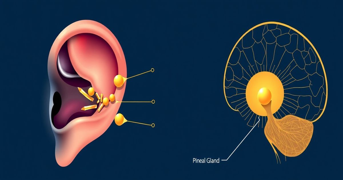
But context matters. Otoconia work because they’re connected to a precisely tuned mechanical system with dedicated neural output. The pineal’s calcite crystals sit in a very different environment. Whether they do anything analogous — in living tissue, in real time — remains an open question.
We don’t know. That’s not hedging — it’s the accurate answer, because nobody has measured it.
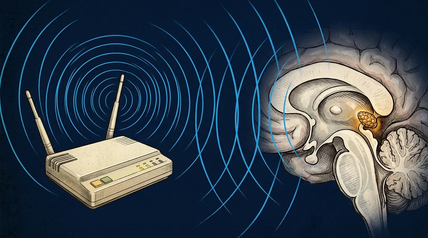
Here’s what was proposed: external electromagnetic fields could interact with the calcite crystals via the inverse piezoelectric effect — fields cause mechanical deformation, deformation stimulates surrounding pinealocytes, pinealocyte stimulation affects signaling cascades including, potentially, melatonin synthesis. The hypothesis runs from outside in, not inside out. It’s mechanistically coherent. Not obviously wrong.
Six years earlier, Lang et al. had already gone looking for exactly this kind of evidence. Their 1996 paper in Bioelectrochemistry and Bioenergetics measured second harmonic generation (SHG) signals from human pineal tissue — a technique that detects non-centrosymmetric crystal structures, which are a prerequisite for piezoelectricity. SHG was positive in all six specimens tested, specifically in pineal tissue, not in adjacent non-pineal brain tissue. That result was what motivated Baconnier’s team to ask: which crystals are producing that signal?
They found calcite.
Actually — let me back up. There’s a complication here that most coverage skips. A 2022 study on otoconia found that SHG signals in those structures are roughly 41 times stronger than in pure calcite alone — and that after removing the calcite with EDTA, the SHG signal actually increased. In otoconia, at least, the signal appears to originate primarily from the organic fibrillar matrix, not the mineral crystals. That doesn’t erase Baconnier’s findings — the calcite is still there, confirmed by four independent methods. But it complicates the inference from “SHG positive” to “piezoelectrically active calcite.” The source of the pineal SHG signal may be messier than the original model assumed.
Twenty-plus years later, no published study has tested the functional hypothesis directly. No cell culture with pinealocytes under applied EM fields. No animal model. No human tissue preparation. The gap isn’t evidence the hypothesis is wrong.
It’s evidence the hypothesis is unfinished.
For a direct analysis of what WiFi and consumer EMF research shows — including the Volkow 2011 JAMA data and calcium channel studies — see does WiFi affect the pineal gland.
I’m going to keep this section tight because it’s a rabbit hole with very little light at the bottom.
The popular version goes: piezoelectric crystals respond to EM fields → stimulate pinealocytes → trigger DMT synthesis → produce altered states. It has the appealing logic of a domino chain. Every piece sounds plausible individually.
On the DMT side: a 2013 study from Barker et al. — working with Jimo Borjigin’s lab at the University of Michigan — detected DMT in microdialysate from the pineal gland of live, free-moving rats using LC/MS/MS. First time that had been done in a living animal. The enzyme required for DMT synthesis, INMT, has been identified in human pineal tissue via immunochemistry.
But none of those links are connected. DMT in rat pineal tissue: documented. INMT in human pineal tissue: documented. Piezoelectric-capable calcite in human pineal tissue: documented. The causal pathway connecting those three facts hasn’t been tested at a single step. Plausible links are not an established chain.
The pineal hypothesis doesn’t exist in isolation. Biological piezoelectricity — the generation of electrical charge from mechanical stress in living tissue — is one of the better-characterized phenomena in biophysics.
The foundational paper came from Fukada and Yasuda in 1957. Working with dry bone samples, they demonstrated that collagen — the structural scaffold bone is built around — generates measurable electrical potentials when mechanically loaded. By the 1970s, researchers had documented piezoelectric responses in cartilage, tendons, dentin, and skin. The field is not fringe. It is textbook biophysics.
Bone remains the most studied example. When bone is bent or compressed, the resulting piezoelectric signal is read by osteoblasts and osteoclasts — the cells responsible for building and resorbing bone. This is why controlled mechanical loading preserves bone density and why prolonged weightlessness causes it to deteriorate. The piezoelectric signal is a feedback loop between mechanical stress and cellular response.
Collagen type I is the operative element. The most abundant protein in the human body, it has a polar triple-helix geometry that creates the non-centrosymmetric arrangement required for piezoelectricity. When deformed, it generates charge. The cell reads the charge. The tissue adapts.
What makes the pineal case different — and harder — is that calcite microcrystals are structurally distinct from collagen. They are inorganic, biomineralized, and their connection to surrounding secretory pinealocytes is unknown. In bone, the electrical signal has a clear cellular target and a well-characterized feedback mechanism. In the pineal, neither the signal pathway nor the cellular target has been identified.
That is not an argument against the hypothesis. It is a description of how far away it is from the mechanistic detail that would make it testable and falsifiable.
Twenty-two years since Baconnier’s paper, the functional question remains untested. That gap is genuinely strange. Here is what proof would require — and why it is harder than it sounds.
In vitro: Isolated pinealocytes cultured in a controlled electromagnetic field environment, with real-time measurement of melatonin output. Sounds straightforward. In practice, primary pineal cell cultures are technically demanding, human pineal tissue is small (100–150 mg in adults), and the experimental design must rule out heating artifacts, cell stress responses, and direct EM effects on melatonin synthesis enzymes — each a confound requiring its own controls.
Ex vivo: Intact pineal tissue under applied EM stimulation, with simultaneous electrophysiological recording and biochemical assay. More informative than cell culture, but the preparation window is narrow and the equipment required spans multiple disciplines.
In vivo: Animal model — most likely rat or sheep — with calibrated EM field application and longitudinal measurement of pineal biomarkers. Most ecologically valid, most expensive, most complex to design. The rat pineal gland has been used in DMT detection studies precisely because it is accessible and well-characterized. That infrastructure already exists in some labs.
Each approach requires different expertise and different funding. A biophysicist who can design the EM stimulation protocol likely does not run a pineal neuroendocrinology lab. The neuroendocrinologist probably cannot design the crystallography controls. The collaboration required cuts across departments and funding streams that rarely overlap.
Negative results would be scientifically valuable — they would close an open question and direct attention elsewhere. But negative results in an underfunded niche rarely generate publications that advance academic careers. The incentives do not favor the experiment getting done.
That is not a conspiracy. It is how scientific resource allocation normally operates.
Two claims. One answer for each.
Claim 1: Piezoelectric crystals are present in the pineal gland. Real. Documented. Baconnier et al. 2002 identified calcite microcrystals with the structural properties associated with piezoelectricity in every specimen examined. Lang et al. 1996 detected SHG signals consistent with non-centrosymmetric crystals in all six pineal tissue specimens. The anatomical finding is solid.
Claim 2: The pineal gland generates or responds to EM fields via piezoelectricity in living humans. Not tested. No functional study exists. This is not the same as “proven false” — it means the experiment hasn’t been done.
Most popular writing collapses these two claims. “The pineal gland is piezoelectric” gets stated as if the gland is actively transducing EM signals right now, while you’re reading this. That’s not a small inferential step. The crystals exist. What they do inside a living system is still unknown.
Why hasn’t this been tested? It’s hard, it’s expensive, and it lives in a no-man’s land between neuroscience, biophysics, and electrophysiology — three fields that don’t naturally collaborate and don’t share funding streams. The topic’s association with spirituality doesn’t help it get taken seriously in grant committees. These are structural problems with how science is funded, not verdicts on whether the question is worth asking.
Let me give you the concrete version.
Baconnier, Lang, and colleagues — working across institutions in France and Israel — published “Calcite Microcrystals in the Pineal Gland of the Human Brain: First Physical and Chemical Studies” in Bioelectromagnetics, volume 23, pages 488–495, PMID 12224052. Three crystal morphologies: cubic, hexagonal, cylindrical. All under 20 micrometers. All calcium carbonate. All structurally distinct from the hydroxyapatite of age-related calcification.
The organic scaffold detected at the ESRF matters. Biomineralized structures — otoconia, bone, shells — form around protein scaffolds. The sulfur signal in Baconnier’s specimens suggests these crystals are the product of a regulated biological process, not incidental mineral deposition.
That’s what makes the twenty-year silence strange. This wasn’t a marginal result in an obscure journal. Bioelectromagnetics is a legitimate peer-reviewed publication. The methodology was rigorous. The paper announced ongoing functional studies. It invited follow-up.
It’s the scientific equivalent of a band releasing a genuinely interesting B-side, announcing a full album, and then going quiet for two decades.

The piezoelectric hypothesis is unconfirmed. Here’s what the science does support, independent of that question.
The evidence on pineal calcification — the hydroxyapatite kind, not the calcite — is more developed. A 2018 review in Molecules by Tan, Xu, Zhou, and Reiter found that in a sample of 346 patients with a mean age of 58.7 years, 62% showed pineal calcification on CT. In a Turkish population study of 12,000 healthy individuals, pineal calcification was the most common form of intracranial calcification, present in 71.6%. The review links progressive hydroxyapatite accumulation to declining melatonin synthesis and downstream health consequences.
That matters independently of piezoelectricity.
Baconnier’s paper noted that the pineal contains two distinct mineral populations with completely different morphologies — and proposed they likely have completely different biological functions. We don’t know what calcite does. We have some evidence on what excess hydroxyapatite may do: compromise the gland’s secretory capacity.
A few things worth tracking:

I’ll be honest: I’m skeptical that “optimizing your pineal crystals” will ever be a meaningful clinical target. The melatonin story is more actionable, better documented, and doesn’t require any of the speculative layers. That’s where I’d put my attention.
None of these require pineal piezoelectricity to be true. They stand on their own evidence.
For more on this, see How to Decalcify the Pineal Gland Naturally, Pineal Gland Calcification Symptoms, and our detailed look at fluoride’s role in pineal health.
Here’s what I actually believe, for whatever that’s worth.
Something real was found in 2002. Calcite microcrystals — biologically synthesized, structurally distinct from the mineral debris of aging, with the crystallographic properties required for piezoelectricity — are sitting in the epithelium of your pineal gland right now. That’s not spiritual speculation. That’s anatomy confirmed by SEM, EDS, SAED, Raman spectroscopy, and synchrotron data.
What nobody knows is what they do.
And the twenty-year gap isn’t reassuring in either direction. It doesn’t mean the hypothesis was disproven — it wasn’t. It means it was proposed, partially supported by a precursor study, published in a legitimate journal with a promise of functional follow-up, and then largely abandoned. That’s a different kind of silence than “this was studied and found to be nothing.”
The practical upshot: you don’t need piezoelectricity to be true to take the pineal gland seriously. The melatonin evidence is real. The calcification data is real. The biological cost of a chronically dysregulated circadian system is well-documented. Start there.
But if you want to keep an eye on the crystal science — the actual peer-reviewed version, not the wellness-influencer version — it’s worth doing. Because if functional piezoelectricity in the pineal gland is ever demonstrated, it will change what we know about how biological tissue interacts with electromagnetic fields.
That paper hasn’t been written yet.
Marcus Hale is an independent researcher and former clinical neuroscientist. The content on PinealCode.com is for informational purposes only and does not constitute medical advice.
4 million results claim iodine decalcifies the pineal gland. The NIS mechanism is real — the extrapolation isn't. Here's where the evidence...

C60 binds up to 6 electrons at once — no other antioxidant works this way. Whether it protects your pineal from calcification: the honest...
WiFi and pineal calcification — what the science actually shows. Volkow 2011, Baconnier 2002 crystals, calcium channels. Honest analysis,...

Marcus Hale
Independent Researcher · Former Clinical Neuroscientist
I spent 12 years in clinical neurology before the questions got more interesting than the answers. PinealCode is where I document what I find at the intersection of brain science and consciousness.